Microscopy & Cell Imaging Core
Olympus BX53 Digital Upright Microscope
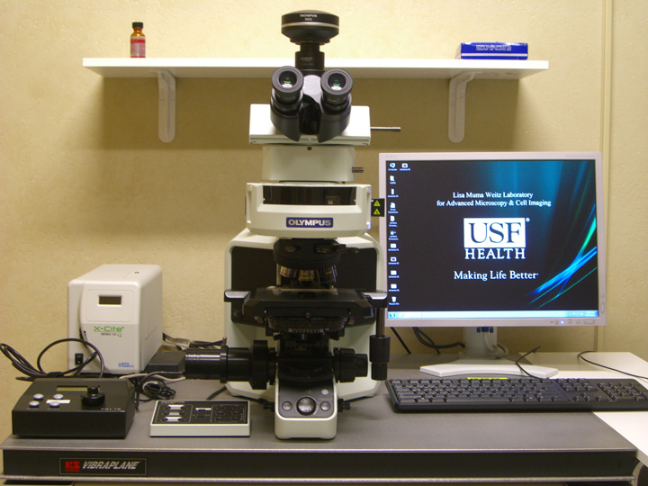
This instrument supports both bright-field and standard fluorescent imaging. It is equipped with a color camera and a monochrome camera. This microscope is capable of capturing bright-field color images with DIC option and of multicolor fluorescence imaging with filters for DAPI, FITC, TRITC and Cy5. It also has a motorized z-axis control for 3D imaging. The CellSense Dimension software provides you with all features necessary to control the microscope and camera and to process and analyze the captured image quantitatively, such as deconvolution, signal intensity, cell counts, volume, surface area and length.
Olympus BX53 and Applications
Olympus BX53
- Lenses:
- 5x MPLAN N 0.10 NA
- 10x UPlanFL N 0.30 NA
- 40x UPlan FL N 0.75 NA
- 60X UPlan FL N 1.25 NA Oil Iris
- Fluorescent filters:
- Violet filter for DAPI, Hoechst
- Green filter for GFP, FITC
- Red filter for RFP, Cy3, TRITC
- Far-red filter for Cy5, DRAQ5
- CCD camera
- Olympus XM10 monochrome camera
- 1.4 Mpixel CCD with 1376 x 1032 pixels (6.45 mm pixel size).
- 14 bit monochrome
- Live frame rate from 15 fps at 1376 x 1032 to 80 fps at 172 x 129.
- Olympus DP26 color comera
- 5 Mpixel CCD with 2448 x 1920 pixels (3.45 mm pixel size).
- 3X12 bit
- Live frame rate from 7 fps at full frame to 31 fps at 320x480.
Applications
This microscope equipped with monochrome and color cameras provides excellent fluorescence and color images. Color camera is very useful for acquiring real color images of histologically or chromogenically stained sample. Monochrome camera with digital pseudocoloring provides multicolor image of fluorescently labeled sample. Indeed, with samples that are about less than 10 mm in thickness, the quality of images is quite comparable to those obtained by confocal microscopy. In case of a thick sample like one shown below (16 mm section of mouse kidney stained with DAPI, Alexa Fluor 488 wheat germ agglutinin, and Alexa Fluor 568 phalloidin), the original captured image may look fuzzy due to light from unfocused area. Deconvolution by cellSense software greatly improves the resolution of the original image.