Microscopy & Cell Imaging Core
Evident Scientific FV1000
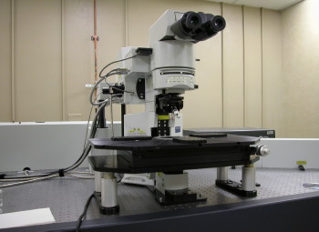
Evident Scientific FV1000 MPE Multiphoton Laser Scanning Microscope
Supports multicolor fluorescent studies for imaging of living, whole mount or thickly sliced specimens. Dynamic biological processes can be imaged hundreds of micrometers within living cells and tissues. Provides support for applications where phototoxicity/photobleaching are a concern such as time course studies of living cells and tissues. Low magnification lens and long working distance stage allow imaging of large samples, embryos, and animals. A range of microprobe objectives are available for minimally invasive in vivo imaging of deep tissues. This microscope also offers conventional confocal laser scanning of samples on slide.
Features & Specification
Features
- Conventional laser scanning fluorescence microscopy of fixed and live samples with visible lasers and DIC imaging.
- Multiphoton laser scanning with Coherent Chameleon Ultra II IR lasers ranging from 680 to 1080 nm.
- Independent grating and slit for precise spectral imaging of dyes with similar emission wavelength.
- Z-series for 3D image.
- Time series for time-lapse imaging (high-speed imaging up to at 16 frames/second)
- FRAP (fluorescence recovery after photobleaching), FRET (fluorescence resonance energy transfer) and photoactivation.
Specification
- Objective lenses
- 4x U Plan APO 0.16 N.A.
- 5x M Plan N 0.10 N.A.
- 10x U Plan APO 0.40 N.A.
- 20x LUC Plan FLN 0.45 N.A.
- 25x XL Plan N 1.05 N.A. Water immersion
- 40x LUM Plan Fl/IR 0.8 N.A. Water immersion
- 40x U Plan FLN 1.30 N.A. Oil immersion
- 60x LUM Plan Fl/IR 0.9 N.A. Water immersion
- 60x U Plan APO 1.42 N.A. Oil immersion
- Fluorescent filters (excitation/emission) for fluorescent viewing.
- DAPI (330-385 nm/420 nm; violet DNA dyes such as DAPI and Hoechst)
- NIBA (470-495 nm/510-550 nm; fluorophores from blue to yellow range, such as CFP, YFP, GFP, Alexa-488, FITC)
- WIG (530-550 nm/ 575 nm for redish fluorophores, like Cy-3, TRITC, DsRED)
- Multiphoton Emission Filter set
- G/R (emission filters; 495-540 nm & 575-630 nm; dichroic 570 nm; suitable for GFP/RFP, FITC/TRITC, Alexa 488/Alexa 594)
- C/Y (emission filters; 460-500 nm & 520-560 nm; dichroic 505 nm; suitable for CFP/YFP)
- V/G (emission filters; 420-460 nm & 495-540 nm; dichroic 485 nm; suitable for DAPI, Hoechst/GFP, Alexa 488, FITC)
Excitation Lasers
| Laser source | Excitation wavelength | Dyes & Fluorophores | Emission color |
|---|---|---|---|
| 405 Diode | 405 nm | DAPI, Hoechst | Violet |
| Multi-line Argon | 457 nm | CFP | Cyan |
| 488 nm | Alexa 488, Oregon Green, FITC, GFP, EGFP, DiO, Cy2 | Green | |
| 514 nm | YFP, EYFP | Yellow | |
| Green HeNe | 543 nm | Cy3, TRITC, mCherry, Alexa 543, Alexa 594 | Orange-red |
| Red HeNe | 633 nm | Alexa 633, Alexa 647, Cy5, TO-PRO3, | Far red |
Fluorochrome Excitation with Multi-Photon IR Laser
| Emission color | Dye | Excitation (FV1000-tested) |
|---|---|---|
| Blue/cyan | Alexa 350 | 780-800 nm |
| Hoechst | 780-800 nm, 900-1100 nm | |
| DAPI | 780-800 nm, 900-1100 nm | |
| CFP | 800-900 nm | |
| Green | Oregon Green | 800-860 nm |
| Alexa 488 | 800-830 nm, (860 nm, 910-920 nm) | |
| EGFP | 920-990 nm | |
| Bodipy | 900-950 nm | |
| FITC | 750-800 nm, (860 nm, 910-920 nm) | |
| DiO | 780-830 nm | |
| Yellow / Orange | YFP | 890-950 nm |
| DiA | 800-860 nm | |
| Red | DiI | 830-920 nm |
| Rhodamine B | 800-860 nm | |
| Alexa 568 | 780-840 nm |
Multiphoton Laser Scanning Image
3T3 cells are stained with DAPI and anti-tubulin (green) antibody and imaged with 800 nm to 920 nm laser to visualize both DAPI and green Alexa fluoro 488 antibody. As seen in the figures, DAPI and Alexa 488 exhibit different excitation and emission profiles over various wavelength; DAPI is excited well with 820~840 nm laser, whereas Alexa 488 shows better exciation with 880~920 nm laser. It is noticeable that some DAPI emissions appear to be detected in Alexa 488 (green) channel at shorter wavelength (i.e. 800 nm), about which one needs to be careful during double immunofluorescence imaging.