Eye Institute
Advanced Visual Function Testing
The experts at USF Health provide visual function testing to determine the most effective prevention, diagnosis, and treatment of diseases affecting the eye and visual system, including retinal diseases and abnormalities of the visual pathways.
Testing for visual function falls into two categories, electrophysiological and psychophysical.
Electrophysiological Tests
-
ERG is a test that measures the changes in the standing potential of the eye induced by brief light stimulation. Many different types of ERG exist (to learn more please follow the Further reading link below). The two tests offered by the USF Eye Institute are named based on the type of light stimulation used.
- Diffuse light: brief (typically 4 msec or less) light stimulus illuminating the whole surface of the retina. The resulting signal and, generally, the test are referred to as flash or full-field ERG. Sometimes this test is called also “standard ERG”, of just “ERG”, as it is the most frequently administered type of ERG test.
- Patterned light: stimulus illuminating the central part of the retina, which is temporally modulated (changes its appearance during the test, typically at least once per second), but is of constant mean luminance. The resulting signal and the test are referred to as pattern ERG (PERG).
What to expect from the test?
Full-field ERG provides information about the functioning of all cell types in the retina. Pattern ERG provides information mostly about the functioning of neurons in the inner retina (e.g. ganglion cells, etc.).How to prepare for the ERG test?
The full-field ERG test lasts about one and a half hours; the pattern ERG test lasts about one hour.Please arrange and plan in advance the mode of transportation to and form the clinic. Full-field ERG typically requires the dilation of both pupils and the effects on your vision can last for several hours. Pattern ERG does not require pupil dilation, but requires distance correction, therefore, it is recommended that you bring your most recent eyeglasses for distance or the equivalent prescription.If you are not sure which test will be performed, please ask our office.
Electrodes will need to be placed on your forehead and earlobe. Please do not apply make-up in the morning and remove all earrings before the test. If you are a contact lens user, please make arrangements to remove and store your contact lenses for the duration of the test (this requirement is valid for both full-field and pattern ERG).
-
Changes in the standing potential of the eye resulting from long-duration (in the order of several minutes) changes in retinal illumination can be recorded indirectly by measuring voltage changes in skin electrodes induced by horizontal movements of the eye following an alternating fixation target. The recording of these changes over time is called electrooculography (EOG). The type of test most frequently used in the clinic typically analyzes the ratio between the decrease in the standing potential in the dark (“dark trough”) vs. the increase of the potential in the light (“light peak”).
What to expect from the EOG test?
The EOG provides information about the functioning of the retinal pigment epithelium and the overlying photoreceptors.How to prepare for the test?
This test takes about 45 minutes. Pupil dilation is not required. -
Electrophysiological signals extracted from the electroencephalographic activity of the visual cortex (recorded from the overlying scalp) as a result of light stimulation are called visual evoked potentials (VEPs). There are several different types of VEP tests. As with ERG, the two types of tests performed at the USF Eye Institute are named based on the type of stimulation used:
- Flash stimulation, and therefore the test is called flash VEP (fVEP)
- When pattern stimulation is used the test is called pattern VEP (pVEP)
What to expect from the test?
Both the flash and the pattern VEP test evaluate the functioning of the optic nerve and the part of the brain responsible for primary analysis of the visual information (“primary visual cortex”). However, the normal functioning of both the optic nerve and the primary visual cortex are depended on a normal functioning of the eye (the retina), therefore, the VEP reflects the overall health of the visual system.How to prepare for the VEP test?
The test takes about one hour.The flash VEP test requires dilation of the pupils, and the effects on your vision can last for several hours, therefore, arrange and plan in advance the mode of transportation to and form the clinic. The pattern VEP does not require pupil dilation, but requires distance correction, therefore, it is recommended that you bring your most recent eyeglasses for distance or the equivalent prescription. If you are not sure which test will be performed, please ask our office.
Electrodes will need to be placed on your head. If you are a contact lens user, you would need to remove temporarily your contact lenses for flash VEP, but not for pattern VEP.
Psychophysical Tests
-
Color vision is the ability of the visual system to distinguish objects based on the wavelengths of the light that reaches the retina. Human's perception of colors is a subjective process whereby the brain responds to the stimuli generated from the cone photoreceptors. The normality of color vision perception can be clinically tested with different methods, including plates (e.g. the Ishihara plates), colored caps (e.g. D-15, Roth 28 hue, FM 100), and other, more elaborate methods (e.g. anomaloscope). Color testing at the USF Eye Institute is based mostly on color cap tests, but can be configured in a different ways, depending on the specifics of the case.
What to expect from the test?
The testing evaluates the ability of the visual system to discriminate between different colors. In addition, some color tests can differentiate between inherited and acquired color deficiencies.
How to prepare for the test?
The testing takes between 30 and 90 minutes, depending on the specific test/s used.
-
In a broad sense, the contrast sensitivity is the ability of the visual system to distinguish different levels of contrast, the contrast being determined by the difference in the color and brightness of the object and other objects within the same field of view. In practical terms it is much easier to test only one of the two elements determining contrast, keeping the other one constant or its contribution reduced to the minimum. In most cases, the color is practically eliminated and the visual system is tested based on its ability to distinguish between the differences in the brightness of an object vs. the surrounding objects (achromatic contrast sensitivity). The testing can be done either by using charts (e.g. Pelli-Robson) or by projecting the stimuli on electronic displays. Currently, the contrast sensitivity testing at the USF Eye Institute is chart-based.
What to expect from the test?
The test evaluates the overall ability of the visual system to detect different levels of contrast. Deficits in this ability can be due to optical factors (e.g. cataract, post-Lasik, etc.), diseases of the inner retina (e.g. glaucoma, diabetic retinopathy, etc.), diseases of the optic nerve (e.g. optic neuritis), drug toxicity (e.g. chloroquine toxicity, vigabatrine toxicity, etc.), or other factors.
How to prepare for the test?
The testing takes between 20 and 45 minutes, depending on the specific test/s used. Distant vision correction (most recent eyeglasses or contact lenses) is required for successful testing. No other preparation is necessary.
-
Other psychophysical visual function tests can include tests for dark adaptation, macular glare tests, etc. At this time, these tests are not offered by the USF Eye Institute.
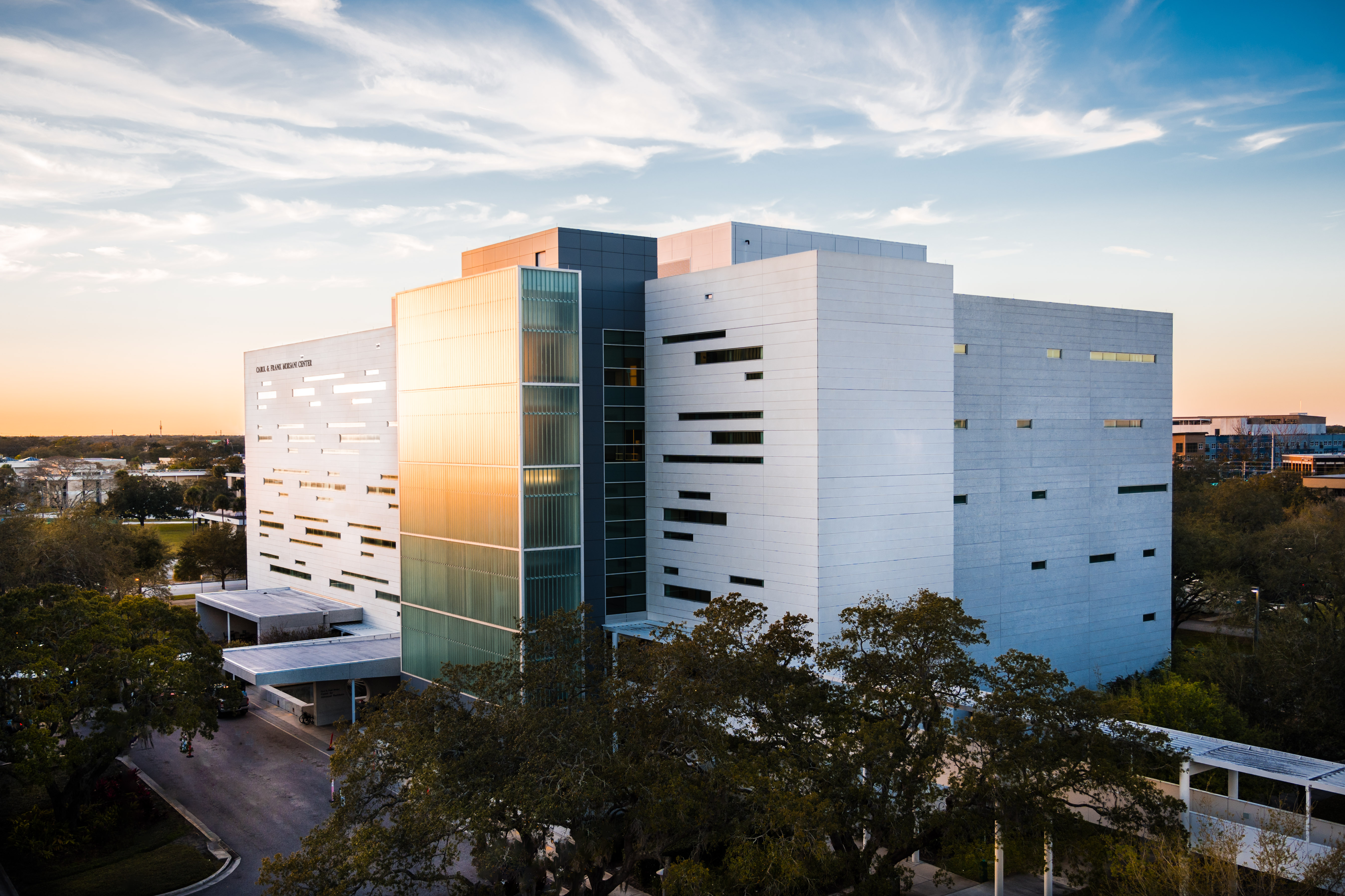
USF Eye Institute at the Carol and Frank Morsani Center for Advance Health Care
Tampa, FL 33612