Laboratory Research
Microscopy Core
The Byrd Institute Microscopy Core serves the needs of research laboratories within the institute and affiliated laboratories. The Microscopy Core aims to provide powerful imaging tools to enable state-of-the-art research on neurological disorders and promote interdisciplinary collaboration.
Dr. Xuewei Wang serves as the managing faculty adviser of the Byrd Microscopy Core. To book, please contact Ajla Kovacs to discuss using this core facility.
Equipment at the Microscopy Core include 6 optical microscope systems with overlapping and unique capabilities:
-
Research Slide Scanner
The SLIDEVIEW™ VS200 research slide scanner captures high-resolution images of stained slides for quantitative analysis. The SLIDEVIEW VS200’s optical system is optimized for scanning sections processed by immunohistochemistry and immunofluorescence. Moreover, the VS200 slide scanner combines five imaging modes in one system with mix-and-match observation method capabilities, so one can find the most appropriate method for their application. Thus, one can switch between brightfield, fluorescence, darkfield, phase contrast, and polarization—or combine different observation methods.
This automated whole slide scanner can also create a virtual library of histology or multi-channel fluorescent slides. Details on the virtual slide can then be zoomed to virtual magnifications from 0.5x-100x and exported as .tif or .jpeg for analysis or presentation with other software packages. The open-source software package QuPath ((multi-platform: Windows, MacOS, Linux) or the free OlyVIA image viewer software from Olympus (Windows only) are the recommended options for processing scanned images.
The VS200 loader has 15 sample trays, each with slots for 6 210 26 × 76 mm (1 × 3 in.) slides. So, one can scan 90 slides in one large batch to finish overnight. You can add additional trays to the loader before all the trays of a given project have been scanned.
-
Airyscan Confocal Microscope
ZEISS LSM 880 (Confocal Laser Scanning Microscope) with Airyscan (1.7X improvement over diffraction limited imaging) provides super resolution imagining, quantitative imaging by linear scanner, imaging of weak fluorescence signals with high sensitivity in AirScan mode. This confocal laser scanning microscope offers high sensitivity, enhanced resolution in x, y and z, and high image-acquisition speed in one system. With airyscan function, it delivers a perfect optical section and superresolution with high sensitivity at 120 nm laterally and 350 nm axially in unique Fast mode speeds.
This imaging system includes the following lasers: 355 nm, 405 nm (UV laser), 440 to 633 nm (VIS laser) and NIR laser, and with the combination of CO2 incubator and temperature control chamber, it can be used for multiple imaging experiments including live cell imaging, FRET and Anisotropy, FRAP and FLIP, FCS and Photoactivation / photoconversion.
-
Multiphoton Confocal Microscope
Nikon’s A1R HD MP, a multiphoton confocal system, features a fast, high resolution galvanometer scanner and an ultra-high speed resonant scanner that is capable of frame rates from 30 fps at 512 x 512 pixels to as fast as 420 fps with both 1024 x 1024 pixel resolution and a large field of view (FOV18), which enables the successful visualization of in vivo rapid changes, such as reactions in living organisms, dynamics and cell interactions.
The multiphoton confocal system with near infrared (NIR) pulse lasers allows investigators to image tissues (i.e. brain) at a subcellular resolution in live animals and ex vivo tissues expressing optogenetic fluorescent proteins or with application of fluorescent probes using up to 5 channels (four-channel episcopic detector plus diascopic detector).
-
Atomic Force Microscope
AFM, a type of scanning probe microscopy, provides very high resolution on the order of fractions of a nanometer, which is more than 1000 times better than the optical diffraction limit. It can be used for imaging of single molecules, live cells, recombinant proteins (i.e. amyoid, tau), or tissues. Measurement outputs can also include elasticity, adhesion, chemical forces and molecular binding sites in physiological conditions. Advantages of using an AFM over TEM include:
- Ease of image interpretation in raster 3D scanning mode (1,000,000X)
- Faster sample preparation time and scanning (5 min) with horizontal resolution of 0.2 nm and a vertical resolution of 0.05 nm
- Ability to function in a liquid environment at physiological conditions, in an air environment or in a vacuum on either conductive or insulating samples
-
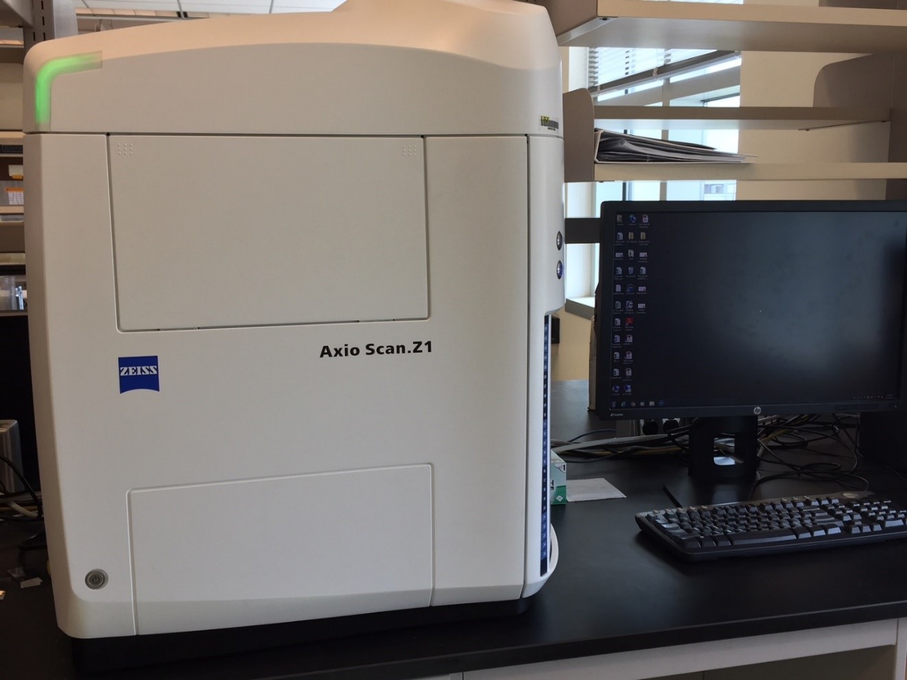
Digital Slide Scanner
ZEISS Axio Scan.Z1, a powerful slide scanner with superior ZEISS optics and automated features, is designed for high throughput brightfield and fluorescence imaging with a high volume quantitative image that is capable of scanning up to 100 whole slides. Images can be acquired using bright-field or captured up to 9 fluorescent channels using standard filter cubes. System resolutions are: 10x - 0.44 µm/pixel, 20x - 0.22 µm/pixel and 40x - 0.11 µm/pixel.
-
Fluorescence Microscope
This microscope, equipped with a Hamamatsu HRm camera and a color AxioCam MRc camera, is an upright, fully motorized, computer controlled high-performance research microscope capable of bright-field, DIC (Differential Interference Contrast) and Fluorescent Imaging by using multiple filter cubes for DAPI, CFP, GFP, YFP, Texas Red and CY5.
-
Automated Microscope
The Leica DM4000 B is an automated microscope for high-end and routine research applications. It comes with a coded 6x or 7x nosepiece, manual z-focus, and motorized fluorescence axis thus providing all basic transmitted-light contrasting methods (brightfield, darkfield, phase, polarization contrast – all fully automated).


Byrd Microscopy Rules
Before using the microscope:
- All users must be trained to use the microscope(s). Only graduate students, post-docs, full-time technicians, and faculty are permitted to use microscopes after training. Please contact Dr. Xuewei Wang to schedule training and to create an account.
Using the microscopes:
- Please start your imaging session punctually (on evenings and weekends as well as regular hours). The charges start automatically from the start of your booked time. Billing will be made in increments of ½ hour.
- Always follow the instructions to turn each microscope on and off as described in the instructions.
- Turn on the microscope computer and login using your name, PI name, and password.
- After finishing the microscopy session, always clean any objectives (oil/water) you used and leave the microscope and room clean. If you bring any live specimens to microscopes, please dispose of them appropriately after imaging.
- You must turn off the microscope computer after your imaging session to indicate the end of your session.
- If you are the last user of the day, make sure to turn off the microscope system according to the specified instructions.
- Please report any problems with the microscopes to Dr. Xuewei Wang.
Canceling:
- If you need to cancel your reservation, please delete your reserved time on the booking calendar BEFORE the booked session.
- If you fail to cancel your reservation and don't use the machine, the charge will still be made.
Image data management:
- Please always transfer your data after you finish imaging. Any images left on the computer hard-drives may be deleted on a regular basis to restore hard drive space.
Byrd Microscopy Core Fee Schedule
| Microscopes | Charges | |
|---|---|---|
| VS200 Whole Slide Scanner | $15/hr | $55/hr |
| ZEISS LSM880 Confocal | $35/hr | $65/hr |
| ZEISS Axio Scan.Z1 | $15/hr | $55/hr |
| JPK NanoWizard 4 Atomic Force Microscope*** | $35/hr | $65/hr |
| Training / Consultation | $25/hr | $65/hr |
***Reusable cantilevers are charged separately ($35-$195/cantilever - depending on application)
Contact Us
For using the whole slide scanner and scheduling a training, please contact Mrs. Ajla Kovacs at abecirbasic@usf.edu
For questions about using microscopes and scheduling a training, please contact Dr. Xuewei Wang at xueweiwang@usf.edu
The 2-photon microscope is not yet available for Core use until further notice