
Animal Imaging Core
Olympus IV 100 Scanning Microscope
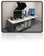 The IV100 Intravital Laser Scanning Microscope has been optimized for collecting light from deep within tissue. Exceptionally high spatial, temporal and multi-wavelength resolution combined with novel far-red and near-IR imaging probes make it possible to image subcellular detail of internal organs and disease processes. With the IV100’s new innovative MicroProbe lenses, researchers are able to do minimal surgery on their animals and yet view the tissue of interest with a powerful objective lens.
Features and Benefits:
- 3 simultaneous detection channels for multicolor fluorescence
- 4 lasers 488, 561, 633, 748 nm
- Immersible MicroProbe lens technology for minimal invasiveness and chemical sterilization
- Multipositional, tilting scanhead
- X,Y,Z,T scan combination
- 76mm range in Z
- Heating plate and anesthesia device
- high resolution 27X magnification (NA 0.7) 3.5 mm-diameter lens with 220 m field of view (FOV).
- high-resolution 20X magnification (NA 0.5) 1.3 mm-diameter lens with 20 m FOV.
- wide-angle 6X magnification (NA 0.14) 1.3 mm-diameter lens with a large FOV of 670 m
- 4X objective lens (NA 0.16)
- 10X objective lens (NA 0.4)
If your experiment requires a surgical procedure, contact Karen Brocklehurst, COM Facility Manager, at 974-9806 or kbrocklehurst@research.usf.edu to reserve a vivarium surgical room and a portable anesthesia machine. Researchers must provide their own isoflurane.
See an hear a PowerPoint training presentation on the Olympus IV 100 Scanning Microscope by Angela Goodacre: click here
For more information visit the Olympus website.